Endra Nexus128 Small Animal Photoacoustic Imaging System introduction Principle of photoacoustic technology: When a beam of light is irradiated onto a biological tissue, the biological tissue absorbs the light energy to generate thermal expansion, and the thermal expansion generates ultrasonic waves, and the amount of absorbed light energy determines the intensity of the generated ultrasonic waves. Different tissues will then produce ultrasound of different intensities, which can be used to distinguish between normal and diseased tissue. Photoacoustic imaging technology detects ultrasonic signals (this technology overcomes the insufficiency of pure optical imaging technology in imaging depth and resolution), reflecting the difference in optical energy absorption (complementing pure ultrasound imaging technology in contrast and function) Sexual defects), combined with the advantages of both optical and ultrasonic imaging techniques, enable high-resolution, high-contrast functional imaging of large depths of tissue. Application field The picture shows tumor vascular system imaging in mice with xenograft tumors. From different angles, the changes in blood vessel diameter, blood vessel distortion, potential blood flow, and blood vessel shunt can be clearly seen. Drug metabolism research: Using molecular imaging technology, real-time monitoring of the movement of labeled drugs in animals, to determine whether the drug can accurately reach the target area and metabolic pathways, as well as therapeutic effects evaluation. Application examples Since the muscles, bones, and proteins of the living organism have a certain degree of absorption in the near-infrared region, as an endogenous contrast agent, different photoacoustic signals can be presented under photoacoustic scanning, and thus can be used as a means of anatomical imaging. . In the photoacoustic imaging of the figure below, we can clearly see the various anatomical structures of the mouse. Â Â Â Â Freeze Dried Powder,Dried Fruit Powder,Freeze Dried Fruit Powder,Freeze Dried Juice Powder Xi'an JCF Herb Technology Development Co., Ltd , https://www.jcfherb.com
--- Powerful preclinical animal disease model imaging tool
Small animal photoacoustic imaging is a new technology with broad prospects in basic research in biomedical research and disease-related applied research. Biomedical research targeting animal models can avoid the risks of experimentation in humans, overcome the shortcomings of certain diseases, long duration, and long duration of disease, and can strictly control animal experimental conditions and reduce the effects of individual differences on imaging. It is one of the indispensable tools in the current animal model research.
Photoacoustic imaging is a non-destructive medical imaging method developed in recent years. It combines the high contrast characteristics of pure optical imaging with the high penetration depth of pure ultrasound imaging to provide high resolution and high contrast tissue imaging. The purchase of this system fully considers the needs of scientific research and practical application, and can carry out cardiovascular diseases (angiogenesis, myocarditis, thrombosis, myocardial infarction, etc.), lymph, tumor, nervous system, blood disease, new molecular exploration for small animals. The cutting-edge research on needle, hemoglobin concentration and blood oxygen saturation measurement and functional imaging will further enhance the research level and status of scientific research units in this field.
Photoacoustic imaging is a landmark advanced molecular imaging research instrument for measuring the scientific research level and depth of scientific research in the fields of life science, basic medicine and chemistry in comprehensive universities. It is currently developing in China and is becoming a key subject in teaching, research and key disciplines. The indispensable analytical test research method for the construction of the room.
The instrument is designed for molecular imaging studies of small animals. Photoacoustic technology has better bio-tissue penetration than near-infrared technology. It also has high resolution and no side effects, and is gradually becoming a non-destructive testing technology for biological tissues. Another research hotspot in the field.
Cardiovascular research: In-depth study of cardiovascular disease (angiogenesis/growth, myocarditis, thrombosis, myocardial infarction, etc.) in small animals, providing quantitative data on hemoglobin concentration and blood oxygen saturation. Such as tumor angiography. Photoacoustic imaging technology can provide high contrast and high resolution images for early detection of tumors, as well as monitoring the treatment process and efficacy of tumors. The rapid growth of tumors is inseparable from the rich vascular system, and studies have shown that tumors are inextricably linked to angiogenesis. Detection of the formation of blood vessels can provide a means of detection for the development of anti-angiogenic drugs, the study of tumor treatment mechanisms, and the establishment of tumor models. 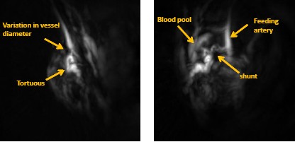
Early diagnosis of disease: Molecular imaging can be used to detect lesions at the molecular level, and to diagnose pathological changes based on pathological changes to achieve early diagnosis of disease.
Observation and study of tumor efficacy: Direct and rapid measurement and tracking of tumor growth and metastasis in various cancer models, and real-time observation and evaluation of changes in hemoglobin concentration and oxygen saturation in cancer treatment.
Gene expression: The expression of genes, cell or tissue specificity, and their response to the environment are observed and studied in living animals.
Stem cells and immune research: real-time observation of stem cell treatment in living animals, and for anti-tumor immunotherapy.
Small animal model: Mark cells, genes, etc. to study the effects of drug treatment in living conditions, such as the treatment of various diseases such as tumors and cardiovascular diseases.
And other applications: such as molecular optics, immunology, nuclear medicine, bone research.
Endra Nexus128 Small Animal Photoacoustic Imaging System Introduction
1. Optical contrast agent application
Many components in our body are endogenous contrast agents. For example, hemoglobin is a good endogenous contrast agent, and hemoglobin carrying oxygen will absorb more at other wavelengths, so according to this principle, light The sound can measure hemoglobin concentration and blood oxygen saturation. The soft tissue in the body and the neovascularization in the tumor are also good contrast agents. The principle of tumor angiogenesis is also based on the very active vascular activity, so the hemoglobin concentration is higher than the internal structure of the tumor, and the tumor can be implemented based on the photoacoustic wavelength of hemoglobin detection. Analysis of angiogenesis.
Exogenous contrast agents, as long as they absorb between 680nm and 950nm, can be detected by photoacoustic imaging systems. For example, commonly used ICGs, almost all nanomaterials, etc., can be produced in photoacoustic systems. Good detection signal.
2. nanomaterials (novel contrast agent) application
Nanomaterials have better light absorption in the near infrared due to their uniform dispersibility and nanometer size, which is also the basis of photoacoustic imaging. Endra Nexus 128, because its excitation wavelength is at 680-950 nm, all nanomaterials include carbon nanotubes, gold nanorods, gold nanocages, and gold nanospheres. Both can have corresponding light absorption at this wavelength, which is very beneficial for the research of novel nanoprobes.
Because the Endra Nexus 128 photoacoustic system features non-invasive detection and because it is true 3-D imaging, it is ideal for continuous observation of experimental animals. After the probe is injected into the experimental animal, the experimental animal can be intermittently scanned to obtain dynamic information that the probe is ingested, absorbed, and cleared in the body.
3. Application in oncology
----- Tumor morphology
Due to its high resolution, photoacoustic can exert its unique advantages in tumor morphology research. At the same time, since photoacoustic detection is a non-invasive and non-destructive detection method, there is no harm to the experimental materials, so the interpretation of the research results is more scientific and reasonable.
---- Tumor perfusion
Due to the different peripheral and internal structures of the tumor, these two different regions cause different behaviors for the absorption of the contrast agent. Peripheral perfusion of tumors is usually faster, because there are more angiogenesis and metabolism, so the clearance rate is fast, and the curve shows a pattern of rapid rise and fall. On the contrary, due to slow metabolism, the perfusion shows slow rise and fall. Mode, and the overall signal peak is much lower than the tumor peripheral signal. This can be used for tumor state determination. If these two peaks are gradually close in time, it indicates that the tumor is inhibited and progresses toward a good prognosis, and vice versa.
---- Probe absorption - dynamic scanning
The absorption analysis of nanoprobes has been described in 4.2. For any commercially available probe and contrast agent, the Nexus 128 can perform a corresponding dynamic scan. Then analyze the ingestion, absorption, and removal processes in the accompanying professional software. The following figure is a dynamic process of absorption and clearance in a tumor model of a mouse using human ICG as an example.
---- Tumor treatment
Since the Nexus 128 can observe tumor morphology and can also detect tumor perfusion and dynamic scanning, it can be applied to the evaluation of tumor treatment. It is possible to directly or semi-quantitatively determine whether the tumor is inhibited from the size of the tumor, or to evaluate the efficacy of the antitumor drug by changes in the internal and external structures of the tumor.
Nexus 128 can also be widely used in other fields of research, such as cardiovascular, drug metabolism, early disease diagnosis, gene expression research, stem cell and immune research, and will not be repeated here.
4. Anatomy application