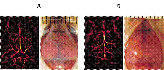
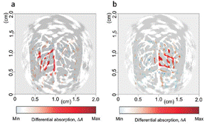
Photoacoustic imaging is a non-destructive medical imaging method developed in recent years. It combines the high contrast characteristics of pure optical imaging with the high penetration depth of pure ultrasound imaging to provide high resolution and high contrast tissue imaging. The small animal photoacoustic imaging system developed by Endra of the United States has a nanomolar sensitivity and a high resolution of 280um to detect the epidermis. Brain imaging and brain function monitoring Photoacoustic imaging proceed brain imaging techniques use is a hot medical imaging technology. Since the optical absorption of brain tissue is closely related to blood oxygen consumption and brain physiological state , photoacoustic imaging can be used to study brain tissue structure and brain function. By monitoring the kinetic changes of cerebral blood oxygen , the dynamic information and functional characteristics of the cranial nervous system can be obtained , which has important application prospects in neurophysiology and neuropathology. At present, the commonly used imaging techniques include brain fMRI (functional magnetic resonance imaging, FMRI) , positive electron emission tomographic scanning techniques (Positron Emission Tomography, PET) and single photon emission computer tomography ( Single Photon Emission Computed Tomography , SPECT) . Compared with the three techniques, photoacoustic technique for brain imaging have not only a non-invasive, lower-cost advantages, but also the distribution characteristics can be obtained oxidized and reduced hemoglobin, providing a more complete the oxygen level of the blood brain distribution portion of the image to be high-level functional activity of the brain to carry out dynamic observation analysis completely without damage situation and to provide high-resolution and high-tissue ratio of the photoacoustic FIG brain Like . Figure 1 Figure 2 Wang et al. applied photoacoustic imaging technology to clearly detect the distribution of cerebral blood vessels in the living mice and the structures of the cerebellum, hippocampus, and lateral ventricles , and obtained clear imaging of brain parenchymal lesions . At the same time, they also obtained the stimulation of mouse beards. An image of cerebrovascular hemodynamic changes in the cerebral cortex center . Figure 2 shows the changes in cerebral vascular hemodynamics in the cerebral cortex center before ( a ) and (b) stimulation of the mouse whiskers . Electro Mechanical Operating Table Neurosurgery Table,Electric Operating Table,Electro Mechanical Surgical Table,Electro Mechanical Operating Table NINGBO TECHART MEDICAL EQUIPMENT CO.,LTD , https://www.techartmed.com
